
A2.2 Cell Structure
A2.2.1 – Cells as Structural Units
Cells are the fundamental building blocks of all living organisms.
All cells have three key structures:
-
Genetic material (DNA)
-
Cytoplasm (mainly water)
-
Plasma membrane (lipids)
A2.2.2 – Microscopy Skills
Microscopes are essential tools in cell biology for visualizing structures not visible to the naked eye.
Types of Microscopes:
-
Light microscope (LM):
-
Uses light and glass lenses.
-
Useful for live specimens and general structure (nucleus, chloroplast, etc.).
-
Limited resolution (~200 nm).
-
-
Transmission Electron Microscope (TEM):
-
Electrons pass through thin sections.
-
High resolution; used to view ultrastructure (e.g., mitochondria, ER, ribosomes).
-
-
Scanning Electron Microscope (SEM):
-
Electrons bounce off the surface.
-
Produces detailed 3D images of cell surfaces.
-
-
Cryo-Electron Microscopy (Cryo-EM):
-
Cells are flash-frozen in their native hydrated state without chemical fixatives.
-
Enables viewing molecular complexes in near-native conditions at atomic-level resolution.
-
Revolutionized the study of macromolecules like ribosomes, viruses, and protein complexes.
-

Magnefication Formula
Magnefication = Image Size/Actual Image
Example:
An image of a cell in a micrograph was measured with a ruler and found to be 60 mm wide. The actual width of the cell is 20 µm. What is the magnification?
Solution:
1. Convert units so they match:
60 mm=60,000 μm
2. Use the formula:
Magnification = Image size/Actual size
Magnification = 60, 000 μm/20 μm= 3000
Magnification = 3000×

A2.2.3 – Developments in Microscopy
dvances in microscopy have enabled scientists to observe cells at increasingly higher resolution and specificity, transforming our understanding of cell structure and function.
1. Fluorescence and Immunofluorescence Microscopy
-
Fluorescent dyes are used to label cellular components; these dyes emit light when excited by specific wavelengths.
-
Immunofluorescence enhances specificity by attaching fluorescent dyes to antibodies that bind to target proteins.
-
These methods allow visualization of specific organelles, proteins, and molecular interactions within cells.
-
Useful for studying both fixed and live cells, including dynamic processes like mitosis or vesicle transport.
2. Freeze-Fracture Electron Microscopy
-
Cells are rapidly frozen and physically fractured, often along the plane of the lipid bilayer.
-
A thin metal replica of the fractured surface is created and observed with TEM.
-
Reveals internal membrane architecture, including integral proteins, pores, and membrane asymmetry.
3. Cryogenic Electron Microscopy (Cryo-EM)
-
Biological samples are vitrified (rapidly frozen in liquid ethane) without chemical fixation.
-
Preserves macromolecules in a near-native, hydrated state.
-
Enables high-resolution 3D reconstruction of complex structures like ribosomes, viruses, and protein complexes.
-
Revolutionized structural biology by allowing atomic-level detail without crystallization.
A2.2.4 – Common Cell Structures
All cells share:
-
DNA as genetic material
-
A cytoplasm where reactions occur
-
A cell membrane regulating exchange
These enable essential life processes.
A2.2.5 - Prokaryotic cell structure

A2.2.6 - Prokaryotic cell structure

A2.2.7 – Processes of Life in Unicellular Organisms
Unicellular organisms carry out all life processes:
-
Metabolism (chemical reactions)
-
Homeostasis (internal balance)
-
Nutrition and Excretion
-
Response to stimuli, Growth, Reproduction
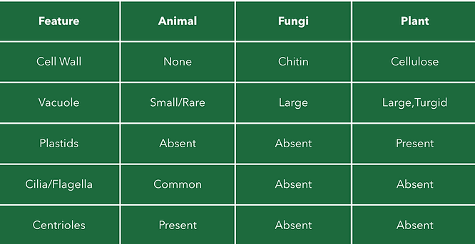
A2.2.8 Animal, Fungal, and Plant Cell Comparison
A2.2.9 – Atypical Eukaryotic Cells
Examples include:
-
Muscle cells – long, multinucleated
-
Sieve tube cells – no nucleus
-
Mammalian red blood cells – lack nuclei
-
Fungal hyphae – no clear cell boundaries

A2.2.10 – Microscopy & Cell Structures
-
Light microscopes show basic structures: membrane, nucleus, chloroplasts.
-
Electron microscopes show detailed ultrastructure: ribosomes, ER, Golgi, mitochondria.
A2.2.11 – Drawing & Annotation from Electron Micrographs
In IB Biology you need to be able to visually understand biological diagrams based on TEM/SEM.
This includes being able to clearly label the structures: nucleus, mitochondria, ER, Golgi, etc.

A2.2.12 – Origin of Eukaryotic Cells by Endosymbiosis
Eukaryotes likely evolved through endosymbiosis:
-
A larger prokaryote engulfed a smaller aerobic prokaryote, which became the mitochondrion.
-
In plants, a second endosymbiosis event involved a photosynthetic prokaryote, forming the chloroplast.
Supporting evidence:
-
Mitochondria and chloroplasts have double membranes, circular DNA, and 70s ribosomes.
-
They replicate independently, like prokaryotes.
Higher Level
A2.2.13 – Cell Differentiation & Specialized Tissues
Multicellular organisms develop specialized tissues through differentiation:
-
All cells contain the same DNA, but different genes are expressed.
-
Gene expression leads to specialization (e.g., nerve cells, muscle cells).
-
Differentiation is guided by chemical signals and position within the embryo.
-
Once differentiated, most cells lose the ability to divide or become other types.
Higher Level
A2.2.14 – Evolution of Multicellularity
Multicellularity evolved as cells formed colonies and became interdependent:
-
Over time, permanent multicellular structures evolved with:
-
Cell specialization
-
Tissue formation
-
Communication systems between cells
Advantages of multicellularity:
-
Larger size, increased complexity
-
Greater efficiency and survival in changing environments